Особенности пробоподготовки образцов монадных форм микроводорослей для сканирующей электронной микроскопии
##plugins.themes.ibsscustom.article.main##
 Google Scholar
##plugins.themes.ibsscustom.article.details##
Аннотация
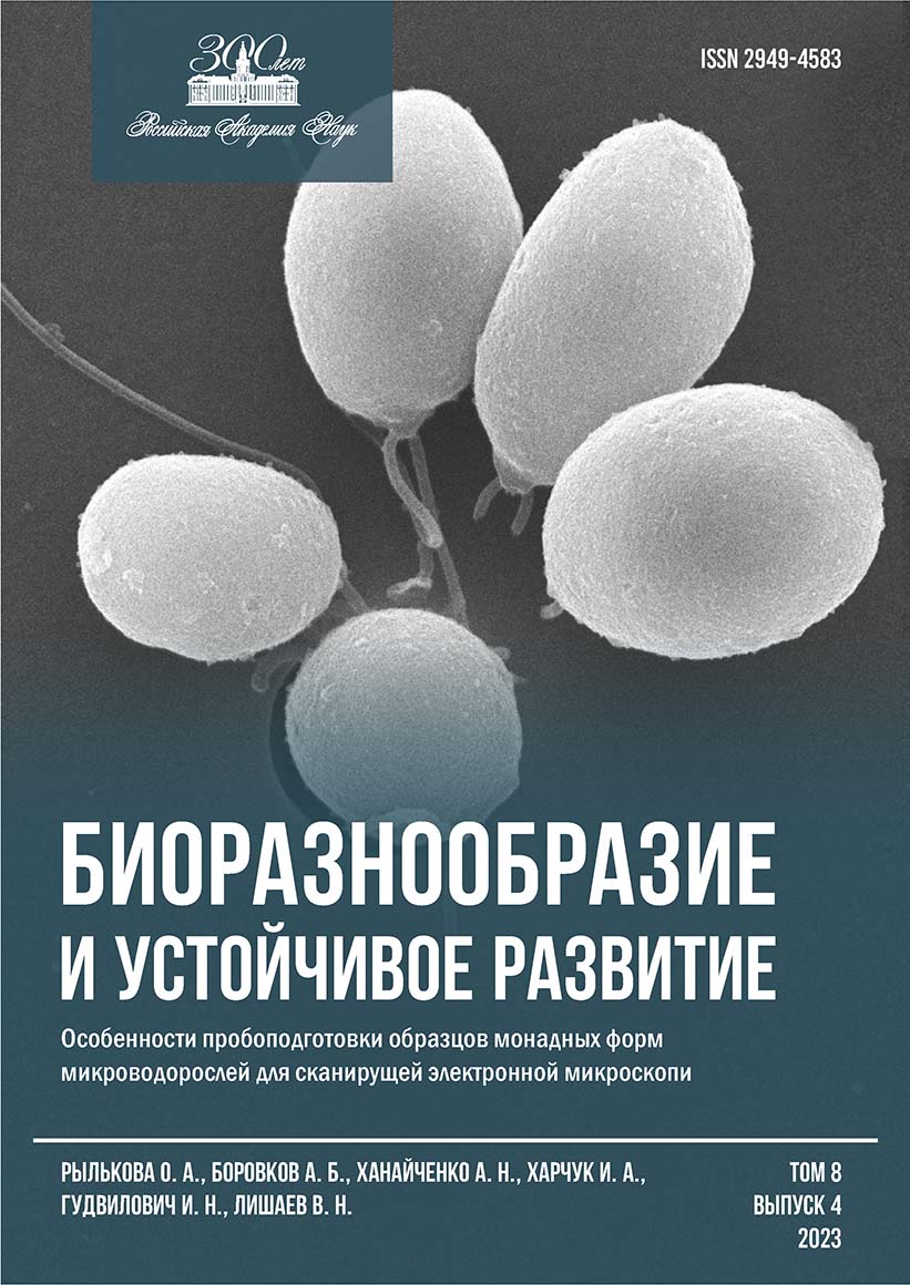
С целью оптимизации пробоподготовки монадных форм микроводорослей для сканирующей электронной микроскопии (СЭМ) проанализированы отечественные и зарубежные руководства. Для отработки методики использовали зелёную микроводоросль Dunaliella salina Teodoresco (штамм IBSS-2 из ЦКП «Коллекция гидробионтов Мирового океана» ФИЦ ИнБЮМ), апробация протокола проведена на криптофитовых водорослях Чёрного моря (Севастопольская бухта). Показано, что при фиксировании материала целесообразно снижать конечную концентрацию (к. к.) глутарового альдегида (глутаральдегида, ГА) в пробе до 1 % (для D. salina) или использовать ступенчатую фиксацию раствором Люголя (для криптофитовых водорослей). При концентрировании микроводорослей, имеющих жгутики, необходимо использовать максимально мягкую фильтрацию (разрежение менее 0,2 атм), промывку пробы проводить только при необходимости, дальнейшую дегидратацию целесообразно осуществлять в бюксе или пластиковом планшете. К хорошему результату приводило использование стёкол, покрытых поли-L-лизином. Показано, что не существует значительной разницы между «этанольной» и «этанольно-ацетоновой» дегидратацией, однако первый способ занимает меньше времени и не требует работы в вытяжном шкафу. Сушка «в критической точке» (2,5–3 ч) и напыление (Au/Pd; 0,5–1,0 мин) соответствовали режимам, обычно рекомендуемым в современных руководствах по пробоподготовке. При невозможности осуществления всех этапов пробоподготовки в один день или в экспедиционных условиях возможно хранение образцов до двух недель в растворе фиксатора или в 75%-ном растворе этанола (в процессе дегидратации). Предложенный протокол предмикроскопной пробоподготовки для исследований с помощью СЭМ может быть использован для изучения поверхностных структур и детализации морфологических характеристик одноклеточных водорослей, имеющих жгутики, и успешно применён при таксономических и биотехнологических исследованиях.
Авторы
Библиографические ссылки
Анисимова О. В. Методы подготовки десмидиевых водорослей (Desmidiales, Charophyta) для изучения в сканирующий электронный микроскоп // Водоросли: проблемы таксономии, экологии и использование в мониторинге : Материалы III междунар. науч. конф., 24–29 авг. 2014 г., Борок / Рос. акад. наук, Ин-т биологии внутр. вод им. И. Д. Папанина. – Ярославль : Филигрань, 2014. – С. 8–10.
Морозова К. Н. Электронная микроскопия в цитологических исследованиях : метод. пособие. – Новосибирск : Изд-во Новосиб. гос. ун-та, 2013. – 85 с.
Algal Culturing Techniques / ed. by R. A. Andersen. – Boston : Elsevier [et al.], 2005. – 578 p.
Bistricki T., Munawar M. A rapid preparation method for scanning electron microscopy of Lugol preserved algae // Journal of Microscopy. – 1978. – Vol. 114, iss. 2. – P. 215–218. – https://doi.org/10.1111/j.1365-2818.1978.tb00131.x
Bratbak G. Microscope methods for measuring bacterial biovolume: epifluorescence microscopy, scanning electron microscopy, and transmission electron microscopy // Handbook of Methods in Aquatic Microbial Ecology / ed. by P. F. Kemp [et al.]. – Boca Raton [et al.] : Lewis Publ., 1993. – P. 309–316. – https://doi.org/10.1201/9780203752746-37
Cerino F., Zingone A. A survey of cryptomonad diversity and seasonality at a coastal Mediterranean site // European Journal of Phycology. – 2006. – Vol. 41, iss. 4. – P. 363–378. – https://doi.org/10.1080/09670260600839450
Clay B. L., Kugrens P., Lee R. E. A revised classification of Cryptophyta // Botanical Journal of the Linnean Society. – 1999. – Vol. 131, iss. 2. – P. 131–151. – https://doi.org/10.1111/j.10958339.1999.tb01845.x
Dolgin A., Adolf J. Scanning electron microscopy of phytoplankton: achieving high quality images through the use of safer alternative chemical fixatives // Journal of Young Investigators. – URL: https://www.jyi.org/2019-july/2019/7/1/scanning-electron-microscopy-of-phytoplanktonachieving-high-quality-images-through-the-use-of-safer-alternative-chemical-fixatives. – Publ. date: July 1, 2019.
Hayat M. A. Principles and Techniques of Electron Microscopy: Biological Applications. – 3rd ed. – [Boca Raton, Florida] : CRC Press, 1989. – 469 p.
Kaufnerova V., Eliaš M. The demise of the genus Scotiellopsis Vinatzer (Chlorophyta) // Nova Hedwigia. – 2013. – Bd. 97, h. 3/4. – P. 415–428. – https://doi.org/10.1127/0029-5035/2013/0116
Khanaychenko A. N., Popova O. V., Rylkova O. A., Aleoshin V. V., Aganesova L. O., Saburova M. Rhodomonas storeatuloformis sp. nov. (Cryptophyceae, Pyrenomonadaceae), a new cryptomonad from the Black Sea: morphology versus molecular phylogeny // Fottea. – 2022. – Vol. 22, iss. 1. – P. 122–136. – https://doi.org/10.5507/fot.2021.019
Murtey M. D., Ramasamy P. Sample preparations for scanning electron microscopy – life sciences // Modern Electron Microscopy in Physical and Life Sciences / ed. by M. Janecek, R. Kral. – Croatia : InTeck, 2016. – Chap. 8. – P. 161–185.
Pomroy A. J. Scanning electron microscopy of Heterocapsa minima sp. nov. (Dinophyceae) and its seasonal distribution in the Celtic Sea // British Phycological Journal. – 1989. – Vol. 24, iss. 2. – P. 131–135. – https://doi.org/10.1080/00071618900650121
Shaish A., Avron M., Ben-Amotz A. Effect of ingibitors on the formation of stereoisomers in the biosynthesis of β-carotene in Dunaliella bardawil // Plant and Cell Physiology. – 1990. – Vol. 31, iss. 5. – P. 689–696. – https://doi.org/10.1093/oxfordjournals.pcp.a077964
Šťastný J., Kouwets F. A. C. New and remarkable desmids (Zygnematophyceae, Streptophyta) from Europe: taxonomical notes based on LM and SEM observations // Fottea. – 2012. – Vol. 12, iss. 2. – Р. 293–313. – https://doi.org/10.5507/fot.2012.021