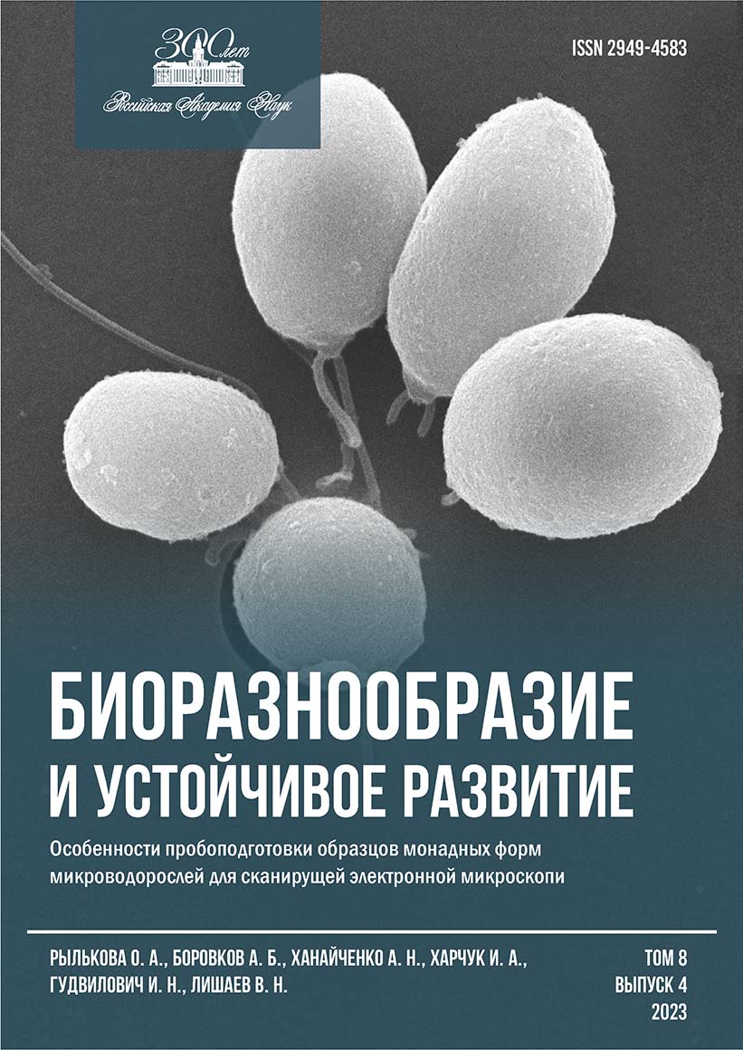
Peculiarities of sample preparation of samples of monadic forms of microalgae for scanning electron microscopy
##plugins.themes.ibsscustom.article.main##
##plugins.themes.ibsscustom.article.details##
Abstract
In order to optimize sample preparation of monadic forms of microalgae for scanning electron microscopy (SEM), domestic and foreign guidelines were analyzed. The green microalga Dunaliella salina Teodoresco (strain IBSS-2 from the Collection of Hydrobionts of the World Ocean, FIC IBSS) was used to develop the technique, and the protocol was tested on the Black Sea (from Collection of the Black Sea cryptophytes, BS-Cry, IBSS). It was shown that it is reasonable reduce the final concentration (f. c.) of glutaric aldehyde (glutaraldehyde, GA) in the sample to 1 % (for D. salina) or to use stepwise fixation with Lugol’s solution (for cryptophytic algae) when fixing the material. When concentrating microalgae equipped with flagella, the mildest possible filtration is necessary (vacuum less than 0.2 atm); the sample should be washed only if necessary; further dehydration should be carried out in a bucket or plastic plate. The use of glasses coated with poly-L-lysine led to good results. It has been shown that there is no particular difference between «ethanol» and «ethanol-acetone» dehydration, but that the former method takes less time and does not require operation in a fume cupboard. The «critical point» drying (2.5–3 h) and sputtering (Au/Pd, 0.5–1.0 min), followed the regimes usually recommended in modern manuals. If it is impossible to carry out all stages of sample preparation in one day or in expeditionary conditions, it is possible to store samples for up to two weeks in fixative solution or in 75 % ethanol solution (in the process of dehydration). The proposed protocol for pre-microscopic sample preparation for SEM studies can be useful for studying surface structures and detailing morphological characteristics of unicellular algae equipped with flagella and has been successfully applied in taxonomic and biotechnological studies.
Authors
References
Анисимова О. В. Методы подготовки десмидиевых водорослей (Desmidiales, Charophyta) для изучения в сканирующий электронный микроскоп // Водоросли: проблемы таксономии, экологии и использование в мониторинге : Материалы III междунар. науч. конф., 24–29 авг. 2014 г., Борок / Рос. акад. наук, Ин-т биологии внутр. вод им. И. Д. Папанина. – Ярославль : Филигрань, 2014. – С. 8–10.
Морозова К. Н. Электронная микроскопия в цитологических исследованиях : метод. пособие. – Новосибирск : Изд-во Новосиб. гос. ун-та, 2013. – 85 с.
Algal Culturing Techniques / ed. by R. A. Andersen. – Boston : Elsevier [et al.], 2005. – 578 p.
Bistricki T., Munawar M. A rapid preparation method for scanning electron microscopy of Lugol preserved algae // Journal of Microscopy. – 1978. – Vol. 114, iss. 2. – P. 215–218. – https://doi.org/10.1111/j.1365-2818.1978.tb00131.x
Bratbak G. Microscope methods for measuring bacterial biovolume: epifluorescence microscopy, scanning electron microscopy, and transmission electron microscopy // Handbook of Methods in Aquatic Microbial Ecology / ed. by P. F. Kemp [et al.]. – Boca Raton [et al.] : Lewis Publ., 1993. – P. 309–316. – https://doi.org/10.1201/9780203752746-37
Cerino F., Zingone A. A survey of cryptomonad diversity and seasonality at a coastal Mediterranean site // European Journal of Phycology. – 2006. – Vol. 41, iss. 4. – P. 363–378. – https://doi.org/10.1080/09670260600839450
Clay B. L., Kugrens P., Lee R. E. A revised classification of Cryptophyta // Botanical Journal of the Linnean Society. – 1999. – Vol. 131, iss. 2. – P. 131–151. – https://doi.org/10.1111/j.10958339.1999.tb01845.x
Dolgin A., Adolf J. Scanning electron microscopy of phytoplankton: achieving high quality images through the use of safer alternative chemical fixatives // Journal of Young Investigators. – URL: https://www.jyi.org/2019-july/2019/7/1/scanning-electron-microscopy-of-phytoplanktonachieving-high-quality-images-through-the-use-of-safer-alternative-chemical-fixatives. – Publ. date: July 1, 2019.
Hayat M. A. Principles and Techniques of Electron Microscopy: Biological Applications. – 3rd ed. – [Boca Raton, Florida] : CRC Press, 1989. – 469 p.
Kaufnerova V., Eliaš M. The demise of the genus Scotiellopsis Vinatzer (Chlorophyta) // Nova Hedwigia. – 2013. – Bd. 97, h. 3/4. – P. 415–428. – https://doi.org/10.1127/0029-5035/2013/0116
Khanaychenko A. N., Popova O. V., Rylkova O. A., Aleoshin V. V., Aganesova L. O., Saburova M. Rhodomonas storeatuloformis sp. nov. (Cryptophyceae, Pyrenomonadaceae), a new cryptomonad from the Black Sea: morphology versus molecular phylogeny // Fottea. – 2022. – Vol. 22, iss. 1. – P. 122–136. – https://doi.org/10.5507/fot.2021.019
Murtey M. D., Ramasamy P. Sample preparations for scanning electron microscopy – life sciences // Modern Electron Microscopy in Physical and Life Sciences / ed. by M. Janecek, R. Kral. – Croatia : InTeck, 2016. – Chap. 8. – P. 161–185.
Pomroy A. J. Scanning electron microscopy of Heterocapsa minima sp. nov. (Dinophyceae) and its seasonal distribution in the Celtic Sea // British Phycological Journal. – 1989. – Vol. 24, iss. 2. – P. 131–135. – https://doi.org/10.1080/00071618900650121
Shaish A., Avron M., Ben-Amotz A. Effect of ingibitors on the formation of stereoisomers in the biosynthesis of β-carotene in Dunaliella bardawil // Plant and Cell Physiology. – 1990. – Vol. 31, iss. 5. – P. 689–696. – https://doi.org/10.1093/oxfordjournals.pcp.a077964
Šťastný J., Kouwets F. A. C. New and remarkable desmids (Zygnematophyceae, Streptophyta) from Europe: taxonomical notes based on LM and SEM observations // Fottea. – 2012. – Vol. 12, iss. 2. – Р. 293–313. – https://doi.org/10.5507/fot.2012.021

 Google Scholar